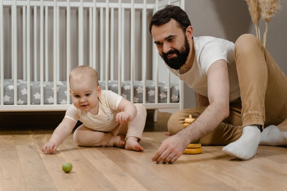
Baby Crossing Eyes: Normal Newborn Vision vs. Strabismus Warning Signs (When to Worry)
Noticed your baby's eyes crossing, wandering, or looking misaligned? Learn the difference between normal infant vision development, pseudostrabismus, and true strabismus warning signs.
Sarah Collins
Mom of Two & Founder of Mommy Tips & Life
Google AdSense Placeholder
Slot ready for "header-banner". Set your AdSense publisher ID in settings when approved.
🩺 Medical Disclaimer: This guide is for educational purposes and should not replace pediatric or obstetric medical consultation.
🛍️ Affiliate Disclosure: We may earn a small commission when you purchase through our links at no extra cost to you.
When my son was around seven weeks old, my husband and I were gazing down at him during tummy time. As he looked up toward a high-contrast black-and-white card, his left eye suddenly darted sharply inward toward his nose while his right eye looked straight ahead. My heart sank into my stomach.
I immediately pulled out my phone and went down a terrifying late-night internet rabbit hole searching terms like “infant crossed eyes permanent blindness” and “baby lazy eye surgery”. The next morning, I rushed him to our pediatrician’s office, practically in tears, holding twenty different smartphone photos capturing his misaligned glances.
Our pediatrician smiled warmly, performed a gentle penlight exam, and immediately put my racing mind at ease. She explained: “Sarah, your baby’s eyes are just learning how to shake hands with each other and talk to his brain.” In the vast majority of cases, intermittent eye crossing in early infancy is a completely normal milestone of neurological vision calibration, or a harmless optical illusion called pseudostrabismus.
Here is the comprehensive medical guide on infant vision milestones, how to spot true strabismus versus optical illusions, and the clinical red flags every parent should monitor.
1. The Timeline of Infant Vision Calibration (0 to 6 Months)
Babies are not born with 20/20 vision or instant stereoscopic depth perception. Visual development is an active neurological learning process that unfolds rapidly over the first six months of life.
Vision Milestones Month by Month:
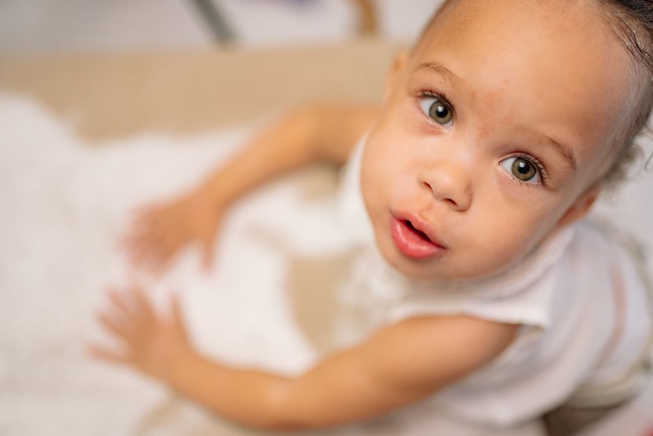
- 0–6 Weeks (The Blurry World): Newborns can only focus clearly on objects 8 to 12 inches away (the exact distance from your chest to your face during feeding). The six extraocular muscles controlling each eyeball lack fine motor coordination, causing eyes to wander, drift, or briefly cross when tired.
- 2–3 Months (Tracking & Social Connection): The brain develops the ability to lock both eyes onto a single moving target. Your baby should begin following a slow-moving toy from side to side and maintaining sustained social eye contact (see our developmental guide on when to worry if a baby is not making eye contact).
- 4–6 Months (Binocular Fusion & Stereopsis): The visual cortex merges the separate images from each eye into one 3D image. Eye wandering should become extremely rare. By 6 months, both eyes should track and focus in seamless unison.
[ 0 to 8 Weeks: Immature Extraocular Muscles ] ➔ Intermittent Wandering / Crossing (Normal) ✅
                                    ⬇
        [ 3 to 4 Months: Visual Cortex Calibrates Binocular Fusion ]
                                    ⬇
    [ 6 Months+: Permanent Straight Eye Alignment & Depth Perception ] 🎯
2. Pseudostrabismus vs. True Strabismus: How to Tell the Difference
One of the most frequent reasons parents panic is Pseudostrabismus (False Strabismus).
Why Your Baby’s Eyes Look Crossed When They Aren’t
Many infants have facial features characteristic of early cranial development:
- A broad, flat nasal bridge.
- Epicanthal folds (extra vertical skin folds covering the inner white corners of the eyes).
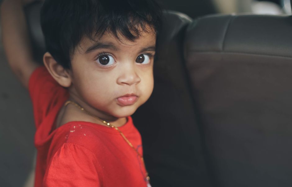
When your baby looks slightly to the left or right, the extra skin fold on the inner corner completely covers the sclera (white part) of the inner eye. Because the white is hidden, the eye appears dramatically turned inward to an observer, even though both pupils are looking in the exact same direction!

The Flashlight Reflection Test (The Hirschberg Test)
Pediatricians use the corneal light reflex to distinguish false crossed eyes from real ocular misalignment:
- Hold a small flashlight or smartphone penlight approximately 2 feet in front of your baby’s face in a dimly lit room.
- Encourage your baby to look directly at the light.
- Observe the pinpoint reflection of white light bouncing off each cornea (pupil).
- In Pseudostrabismus (Normal Alignment ✅): The pinpoint white light reflection hits the exact dead center of both pupils simultaneously.
- In True Strabismus (Misalignment ⚠️): The light reflection is centered in one pupil, but displaced toward the outer edge (in esotropia/inward turn) or inner edge (in exotropia/outward turn) of the other pupil.
3. The 4 Types of True Strabismus
When true ocular misalignment occurs, it is categorized by the direction in which the deviating eye turns:
          [ Esotropia ] ────────── Eyes Turn INWARD (Crossed)
          [ Exotropia ] ────────── Eyes Turn OUTWARD (Wall-Eyed)
          [ Hypertropia ] ──────── One Eye Drifts UPWARD
[ Hypotropia ] ───────── One Eye Drifts DOWNWARD- Infantile Esotropia: The most common form, where one or both eyes turn inward toward the nose. It typically presents before 6 months of age.
- Accommodative Esotropia: Often appears between ages 1 and 3 when a child tries to focus hard on close objects to overcome farsightedness (hyperopia).
- Exotropia: The eye drifts outward toward the ear, especially when the child is fatigued, daydreaming, or looking into bright outdoor sunlight.
- Vertical Strabismus (Hyper/Hypotropia): Rare conditions where one eye rests higher or lower than the other.
4. Why Early Detection Is Critical (Preventing Amblyopia)
The human brain is remarkably adaptable. If one eye is misaligned, the brain receives two conflicting images (double vision). To prevent disorientation, the infant’s visual cortex will actively suppress and ignore the image coming from the misaligned eye.
[ Misaligned Eye Sends Blurry/Offset Image ] ➔ [ Brain Suppresses Visual Input from Weak Eye ]
                                    ⬇
    [ Neural Pathways Fail to Develop ➔ "Lazy Eye" (Amblyopia) / Permanent Vision Loss ] ⚠️
If left untreated during the critical developmental window (before age 6 to 7), the suppressed eye may lose permanent visual acuity. When caught early, strabismus can be effortlessly corrected with corrective glasses, temporary occlusion patches over the strong eye to strengthen the weaker neural pathway, or minor muscle realignment surgery.
To support healthy early cognitive and sensory tracking without visual overstimulation, ensure you are avoiding overstimulating neon sensory cartoons and screen time traps.
5. Diagnostic Comparison: Normal Eye Calibration vs. Strabismus Warning Signs
Use this diagnostic table to evaluate your baby’s visual behavior:
| Visual Behavior | Normal Developmental Phase 🌿 | Strabismus / Red Flag ⚠️ |
|---|---|---|
| Frequency of Crossing | Intermittent (a few seconds when tired or nursing) | Constant (eye is turned in or out 100% of the time) |
| Age of Occurrence | Common from 0 to 3 months | Persistent past 4 to 6 months of age |
| Corneal Light Reflection | Symmetrical, centered in both pupils (Hirschberg test) | Asymmetrical, displaced in one pupil |
| Head Posture | Holds head centered when looking at toys | Tilts head or turns face to one side to compensate for double vision |
| Pupil Color in Photos | Symmetrical red/orange reflex in both eyes | Asymmetrical white, yellow, or absent reflex (Leukocoria) |
| Eye Movements | Smooth tracking by 3–4 months | Jerky, rapid, involuntary shaking movements (Nystagmus) |

Pediatric Red Flags: When to Call the Doctor
Contact your pediatrician or request a referral to a Pediatric Ophthalmologist if you notice any of the following:
⚠️ Warning Alert: Immediate Evaluation Needed
- Constant Eye Turn: One eye is turned in, out, up, or down at all times, even during alert morning play.
- Lack of Fixation by 3 Months: Your baby does not look at your face or follow moving objects held in front of them (see our guide on baby not responding to social cues and milestone signs).
- White Pupil Reflex (Leukocoria): In flash photography or dim light, one pupil looks white, silvery, or cloudy instead of the standard red reflex. This requires emergency evaluation to rule out congenital cataracts or retinoblastoma.
- Persistent Eye Rubbing & Light Sensitivity: Squinting or closing one eye tightly whenever stepping outdoors in daylight.
Simple Floor Play Activities to Support Visual Tracking
To encourage strong binocular coordination during your daily routine:
- High-Contrast Card Tracking (0–8 Weeks): Slowly move a black-and-white patterned card in an arc 10 inches from your baby’s face, pausing to let them lock their gaze.
- Unrestricted Floor Tummy Time: Floor play strengthens the neck, shoulder, and back muscles required for stable head and eye coordination (and prevents issues like container baby syndrome).
- Face-to-Face Sing-Alongs: Your face is your baby’s favorite visual stimulus. Make exaggerated, playful facial expressions while moving your head slowly from side to side.
Remember, seeing your newborn’s eyes briefly cross during a sleepy burp is almost always just a sign of a growing nervous system learning the ropes. Stay observant, take comfort in the developmental timeline, and trust your maternal instincts!
Google AdSense Placeholder
Slot ready for "in-article". Set your AdSense publisher ID in settings when approved.
Frequently Asked Questions by Moms
Is it normal for a newborn baby's eyes to cross occasionally? ▼
Yes, intermittent eye crossing or wandering is completely normal in newborns and infants up to 3 to 4 months of age. In the early weeks, the six extraocular muscles controlling eye movement and the neural pathways connecting the brain to binocular vision are still maturing.
At what age should a baby stop crossing their eyes? ▼
By 4 months of age (and certainly by 6 months), an infant's eyes should work together in coordinated binocular alignment and track moving objects smoothly without drifting inward or outward.
What is pseudostrabismus (false crossed eyes) in babies? ▼
Pseudostrabismus is an optical illusion where a baby's eyes appear crossed due to a wide, flat nasal bridge and prominent skin folds in the inner corners of their eyelids (epicanthal folds). While the eyes look crossed to the naked eye, the corneal light reflection is perfectly centered in both pupils.
When should I take my baby to see a pediatric ophthalmologist? ▼
You should schedule a pediatric ophthalmology evaluation if your baby's eyes are constantly crossed (at all times), if one eye is persistently turned inward or outward past 4 months of age, if their eyes make rapid involuntary shaking movements (nystagmus), or if you notice a white glow in their pupil in flash photos (leukocoria).

Written by Sarah Collins
Doctor-Reviewed & Fact-CheckedMom of Two & Founder of Mommy Tips & Life
Sarah is a mother of two (Liam & Maya) sharing evidence-based parenting guides, infant sleep solutions, and baby safety alerts tested in real family life and vetted against AAP clinical standards.
Google AdSense Placeholder
Slot ready for "footer-banner". Set your AdSense publisher ID in settings when approved.